 | ||||
Bacterial Cell Wall Structure
Location & Amount of Peptidoglycan in
Gram-positive vs. Gram-negative Bacteria
Peptidoglycan (pep-tid-o-gly-can) is a molecule found only in the cell walls of bacteria. Its rigid structure gives the bacterial cell shape, surrounds the plasma membrane and provides
prokaryotes with protection from the environment.
Article Summary: Amount and location of the peptidoglycan molecule in the prokaryotic cell wall determines whether a bacterium is Gram-positive or Gram-negative.
Bacterial Cell Wall Structure: Gram + & Gram -
You have free access to a large collection of materials used in two college-level introductory microbiology courses (8-week & 16-week). The Virtual Microbiology Classroom provides a wide range of free educational resources including PowerPoint Lectures, Study Guides, Review Questions and Practice Test Questions.
Gram-positive
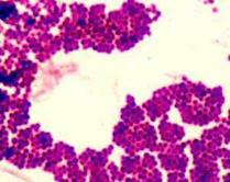
Staphylococcus. Thick peptidoglycan cell wall retains crystal violet primary Gram stain. Go to >
Page last updated 3/2016
 | ||||||
SPO VIRTUAL CLASSROOMS
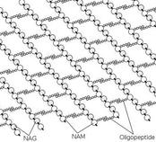
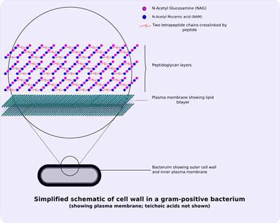
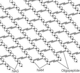
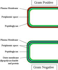
Peptidoglycan is a huge organic polymer; a mesh-like series interlocking strands of sugars -- N-acetylglucosamine (NAG) and N- acetylmuramic acid (NAM) -- cross-linked by short amino acid bridges.
Gram-positive Cells
In Gram-positive bacteria, peptidoglycan makes up as much as 90% of the thick cell wall enclosing the plasma membrane.
SPO VIDEO:
How to Do a
Gram Stain
During Gram staining, these thick, multiple layers (20–80 nm) of peptidoglycan retain the dark purple primary stain crystal violet, whereas Gram-negative bacteria stain pink.
